Key Takeaways & Executive Findings
- •• SDT combines low-intensity ultrasound and sonosensitizers to generate cytotoxic ROS, offering a non-invasive approach with deep tissue penetration for treating deep-seated tumors. • Nanomaterials enhance SDT by improving sonosensitizer delivery, tumor accumulation, and overcoming hypoxia, thereby increasing therapeutic efficacy. • The review highlights recent advances in nanosonosensitizers for both monotherapy and synergistic therapy, particularly for orthotopic tumors that are challenging for conventional treatments. • Despite promising preclinical results, the unclear mechanism and suboptimal efficiency of SDT remain barriers to clinical translation, necessitating further research.
Abstract
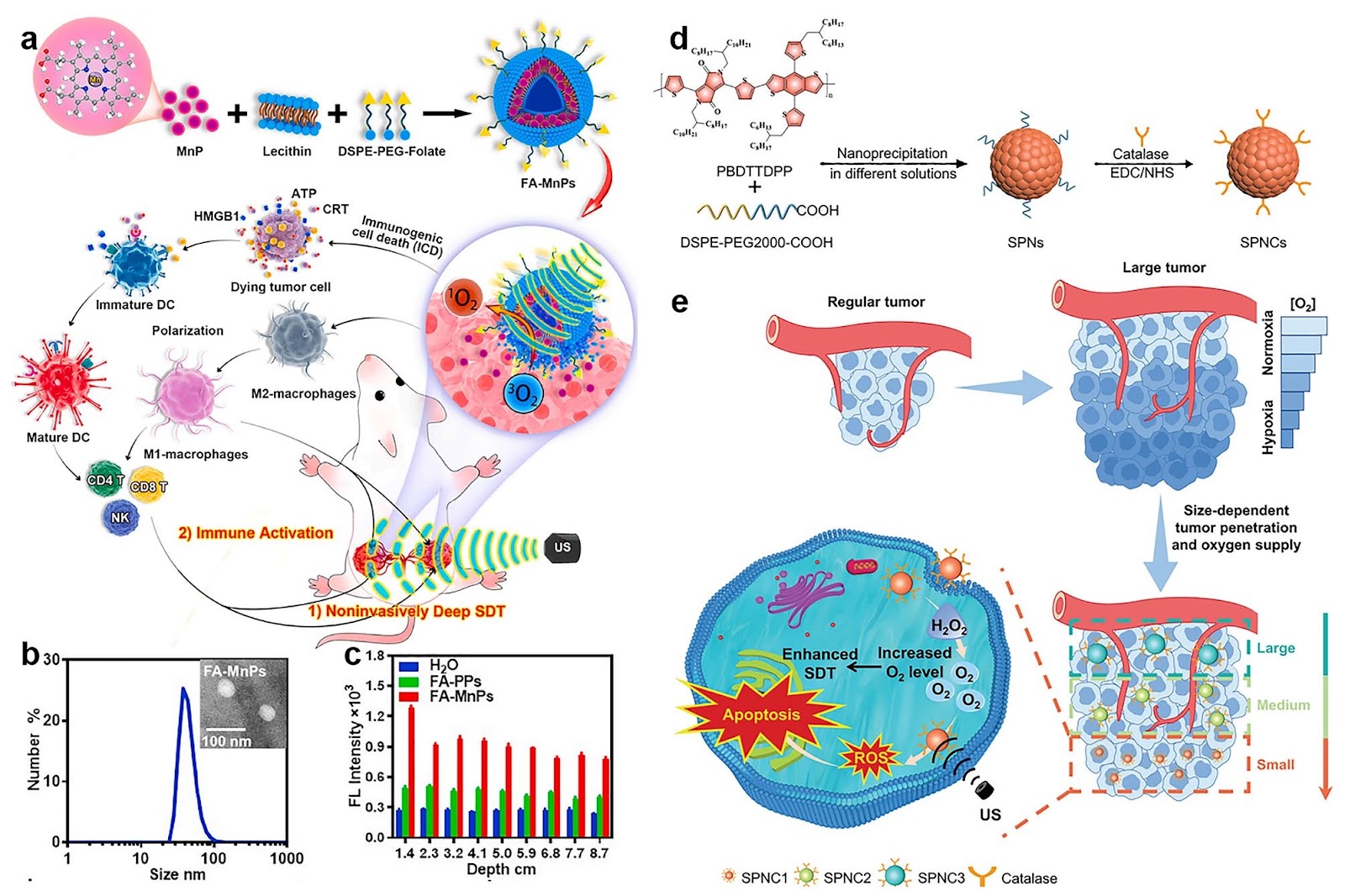
Sonodynamic therapy (SDT) as an emerging modality for malignant tumors mainly involves in sonosensitizers and low-intensity ultrasound (US), which can safely penetrate the tissue without significant attenuation. SDT not only has the advantages including high precision, non-invasiveness, and minimal side effects, but also overcomes the limitation of low penetration of light to deep tumors. The cytotoxic reactive oxygen species can be produced by the utilization of sonosensitizers combined with US and kill tumor cells. However, the underlying mechanism of SDT has not been elucidated, and its unsatisfactory efficiency retards its further clinical application. Herein, we shed light on the main mechanisms of SDT and the types of sonosensitizers, including organic sonosensitizers and inorganic sonosensitizers. Due to the development of nanotechnology, many novel nanoplatforms are utilized in this arisen field to solve the barriers of sonosensitizers and enable continuous innovation. This review also highlights the potential advantages of nanosonosensitizers and focus on the enhanced efficiency of SDT based on nanosonosensitizers with monotherapy or synergistic therapy for deep tumors that are difficult to reach by traditional treatment, especially orthotopic cancers.
1. Introduction
Nowadays, cancer has become one of the main causes of human death, and the prevalence rate is still on the rise every year [1–4]. Although the existing three major treatments, including surgery, chemotherapy, and radiotherapy, have achieved certain results in the treatment of cancer, serious adverse effects on normal tissues are inevitable for chemotherapy and radiotherapy as well as traumatic for surgery [5, 6]. Thus, it is urgent to develop an effective and precise non-invasive treatment. With the development of science and technology, phototherapy, including photodynamic therapy (PDT) and photothermal therapy (PTT), has developed a lot [7–9]. During PDT and PTT, the core component photosensitizer is excited under light irradiation with a suited wavelength and produces reactive oxygen species (ROS) and heat to kill tumor cells, respectively [10–14]. Phototherapy has a good application prospect owing to the merits of spatiotemporal selectivity, non-invasiveness, non-drug resistance, and less side effects [15–20]. However, because of the poor penetration depth of light, phototherapy is limited to the cure of lesion on the skin or surface [21–23].
In contrast, ultrasound (US) as one kind of mechanical waves has stronger tissue penetration, which is widely applied in the diagnosis of abdominal diseases [24, 25]. In recent years, it has made great progress in the fields of cardiac US, obstetrics and gynecology US, and endovascular US, so it has shown extensive application potential in the detection and treatment of deep-level tumors [26, 27]. In 1989, Umemura et.al found that the combination of US irradiation and hematoporphyrin showed effective antitumor effects in vivo and in vitro, which was attributed to the hematoporphyrin and was activated by US cavitation and generated the cytotoxic ROS [28]. By 1992, Umemura et al. proposed that this treatment was called “sonodynamic therapy” [29, 30]. As an emerging type of non-invasive modality, sonodynamic therapy (SDT) is developed based on PDT, mainly involving low-intensity US and sonosensitizers and having similar principles and function [31, 32]. Compared with other non-invasive treatment such as PDT and PTT, US possesses low tissue attenuation and deep penetration, allowing it to reach tissues up to more than 10 cm in depth [33]. This is significantly greater than the ≈1 cm penetration depth of near-infrared (NIR) light within tissues [34]. Consequently, SDT can address the limitation of traditional phototherapy in treating deep tumors. Furthermore, US is clinically recognized as a safe and effective imaging modality [35]. Hence, SDT holds great promise for clinical applications.
Loading authentic research manuscript (Pages 1–5)...
Mengyao Yang, Xin Wang, Mengke Peng, Fei Wang, Senlin Hou, Ruirui Xing, Aibing Chen (2025). Nanomaterials Enhanced Sonodynamic Therapy for Multiple Tumor Treatment. Nano-Micro Letters. https://doi.org/10.1007/s40820-025-01666-8
Research & Educational Purpose Only:The translations, structured abstracts, analytical annotations, and data reports provided by SinoTechIntel are intended exclusively for academic research, internal corporate R&D, and educational benchmarking. They do not constitute formal engineering, chemical safety, legal, or professional advice.
Copyright & Intellectual Property Notice: Original copyright of the underlying source articles and experimental data remains with the respective authors, institutions, and original publishing journals. SinoTechIntel claims intellectual property only over its proprietary translations, analytical syntheses, and AEO structured enhancements in accordance with international fair use and academic citation principles.
Frequently Asked Questions
What is sonodynamic therapy (SDT)?
Sonodynamic therapy (SDT) is an emerging non-invasive cancer treatment that combines low-intensity ultrasound and sonosensitizers to generate cytotoxic reactive oxygen species (ROS) that kill tumor cells. It offers deep tissue penetration and minimal side effects compared to traditional therapies.
How do nanomaterials enhance sonodynamic therapy?
Nanomaterials enhance SDT by improving the delivery and accumulation of sonosensitizers at tumor sites, overcoming tumor hypoxia, and enabling synergistic therapies. They can be engineered to respond to ultrasound, increasing ROS production and therapeutic efficacy.
What are the main challenges of SDT?
The main challenges of SDT include the unclear underlying mechanism, suboptimal therapeutic efficiency, and limited clinical translation. Additionally, the development of efficient and biocompatible sonosensitizers remains a hurdle.
What types of sonosensitizers are used in SDT?
Sonosensitizers are classified into organic (e.g., porphyrins, phthalocyanines) and inorganic (e.g., titanium dioxide, gold nanoparticles) types. Nanosonosensitizers, which are nanomaterial-based, are being developed to improve solubility, targeting, and ROS generation.
Why is SDT particularly promising for deep tumors?
Ultrasound can penetrate tissues up to 10 cm or more, significantly deeper than light used in phototherapy (about 1 cm). This allows SDT to treat deep-seated tumors, such as orthotopic cancers, that are difficult to reach with conventional non-invasive methods.
Related Technical Papers & Translations
Direct Repair of the Crystal Structure and Coating Surface of Spent LiFePO4 Materials Enables Superfast Li-Ion Migration
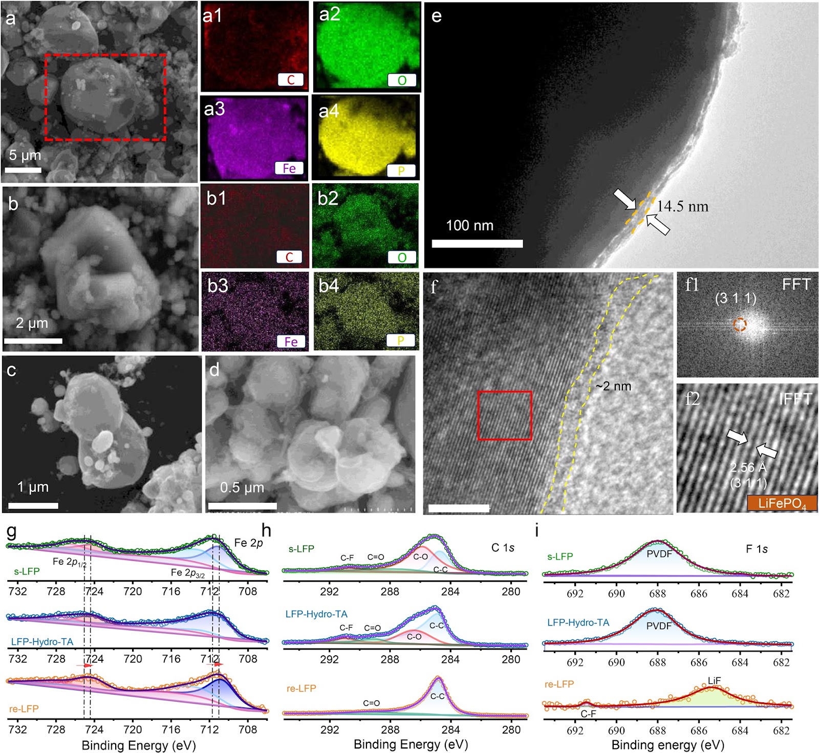
The rapid accumulation of spent LiFePO4 (LFP) cathodes from retired lithium-ion batteries necessitates the development of effective and environmental-friendly recycling strategies. In this context, direct regeneration has emerged as a promising approach for reclaiming LFP cathode materials, offering a streamlined pathway to restore their electrochemical functionality. We report an integrated regeneration protocol that simultaneously repairs the degraded crystal structure and reconstructs the damaged carbon coating in spent LFP. The regenerated cathode material had superfast lithium-ion diffusion kinetics and a stable cathode–electrolyte interface, giving a remarkable rate capability with specific capacities of 122 mAh g−1 at 5C and 106 mAh g−1 at 10C (1C = 170 mA g−1). It also maintained capacities of 110.7 mAh g−1 (5C) and 84.1 mAh g−1 (10C) after 400 cycles. It could be used in harsh environments and could be stably cycled at subzero temperatures (−10 and −20 °C) and in solid-state electrolyte batteries. Life cycle assessment combined with economic evaluation using the EverBatt model reveals that this direct regeneration approach has high economic and environmental benefits.
Oxide Semiconductor for Advanced Memory Architectures: Atomic Layer Deposition, Key Requirement and Challenges
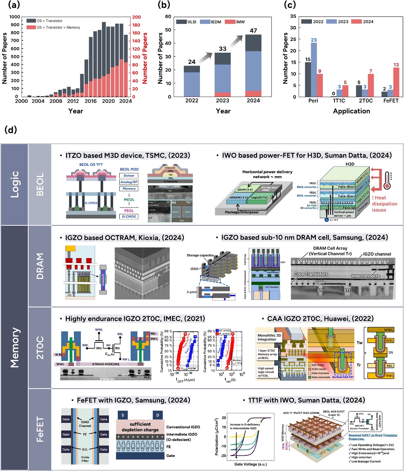
Oxide semiconductors (OSs), introduced by the Hosono group in the early 2000s, have evolved from display backplane materials to promising candidates for advanced memory and logic devices. The exceptionally low leakage current of OSs and compatibility with three-dimensional (3D) architectures have recently sparked renewed interest in their use in semiconductor applications. This review begins by exploring the unique material properties of OSs, which fundamentally originate from their distinct electronic band structure. Subsequently, we focus on atomic layer deposition (ALD), a core technique for growing excellent OS films, covering both basic and advanced processes compatible with 3D scaling. The basic surface reaction mechanisms—adsorption and reaction—and their roles in film growth are introduced. Furthermore, material design strategies, such as cation selection, crystallinity control, anion doping, and heterostructure engineering, are discussed. We also highlight challenges in memory applications, including contact resistance, hydrogen instability, and lack of p-type materials, and discuss the feasibility of ALD-grown OSs as potential solutions. Lastly, we provide an outlook on the role of ALD-grown OSs in memory technologies. This review bridges material fundamentals and device-level requirements, offering a comprehensive perspective on the potential of ALD-driven OSs for next-generation semiconductor memory devices.
Laser powder bed fusion of biodegradable Zn-4Cu alloy: Processing, microstructure and properties
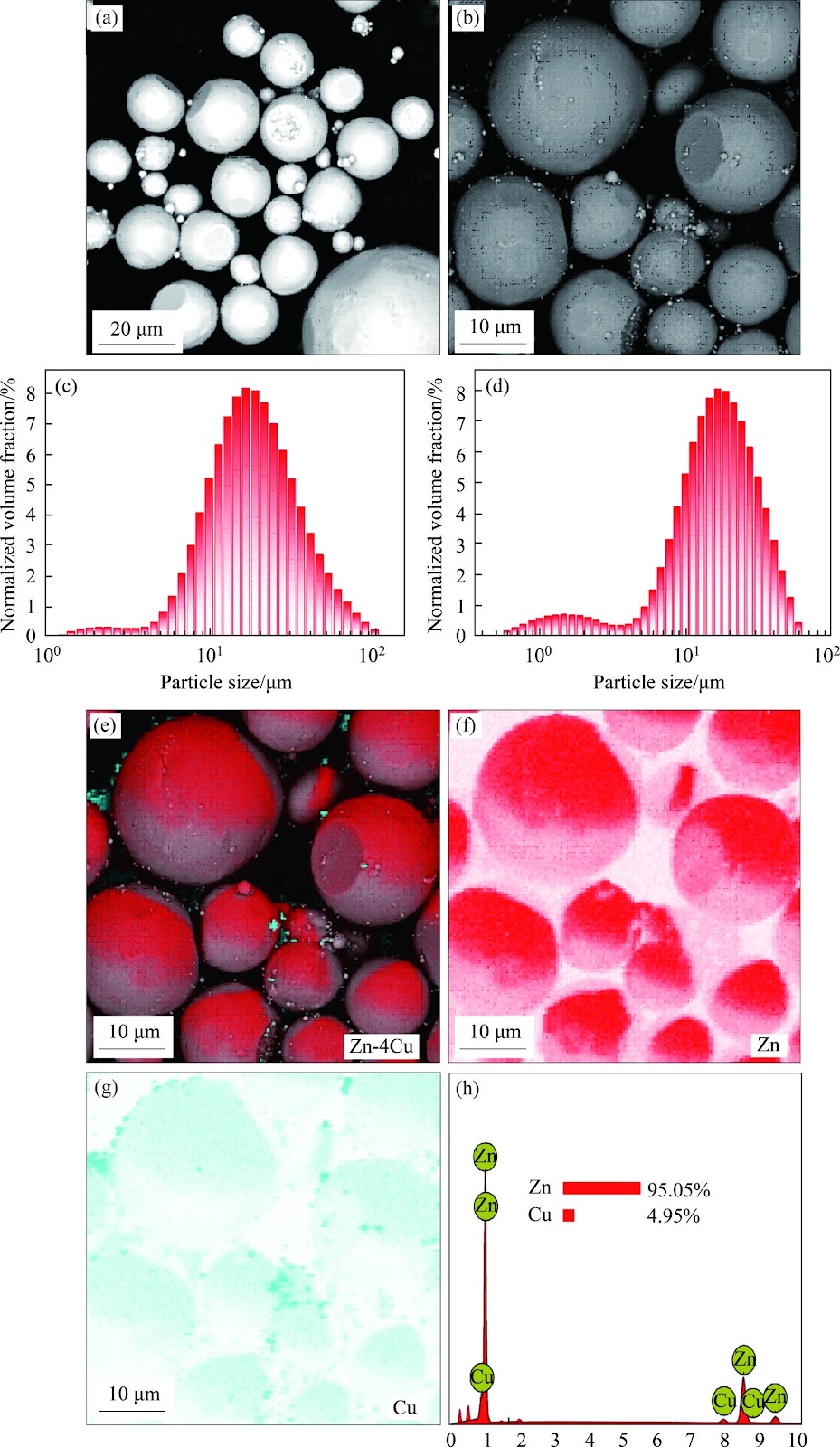
Zn's natural degradability and biocompatibility make it a promising candidate for implants, however, its mechanical properties remain insufficient for bone applications. In this study, the performance of Zn was enhanced by developing Zn-Cu alloys via laser powder bed fusion (LPBF). Optimal LPBF parameters for forming stable tracks were achieved by adjusting laser power and scanning speed. Under optimized conditions of 100 W and 100 mm/s, high-density (99.58%) Zn-Cu alloys with improved hardness (68.2HV) and yield strength (160 MPa) were achieved. These improvements are attributed to solid solution strengthening, segregation strengthening, and grain refinement. The Zn-Cu alloys also demonstrated favorable degradation behavior, with a rate of 0.16 mm/year. This degradation is primarily driven by micro-galvanic corrosion between the CuZn5 phase and Zn matrix, along with refined grains and increased grain boundary density. This work demonstrates a viable strategy for fabricating Zn-based implants with enhanced structural integrity and mechanical performance via LPBF.