Key Takeaways & Executive Findings
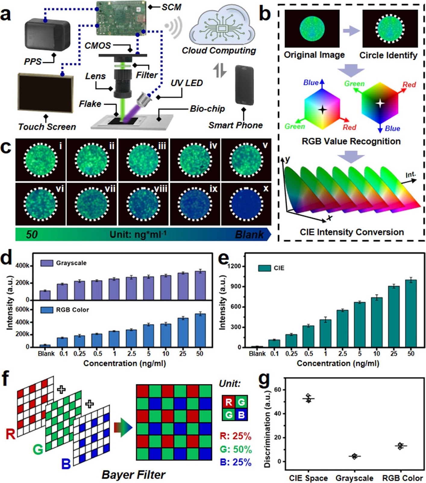
- •• A novel intelligent biosensing platform integrates quantum dots luminescence, microfluidic biochip, and machine vision algorithm for point-of-care CEA protein diagnostics. • The platform achieves an outstanding detection limit of approximately 0.021 ng mL−1 for CEA, surpassing commercial lateral flow assay strips. • Machine vision algorithm enhances portability and integration, expanding the potential of point-of-care biosensing applications. • Validation with human-sourced artificial saliva samples demonstrates superior detection capabilities compared to commercial rapid test strips.
Abstract
Realizing the point-of-care tumor markers biodetection with good convenience and high sensitivity possesses great significance for prompting cancer monitoring and screening in biomedical study field. Herein, the quantum dots luminescence and microfluidic biochip with machine vision algorithm-based intelligent biosensing platform have been designed and manufactured for point-of-care tumor markers diagnostics. The employed quantum dots with excellent photoluminescent performance are modified with specific antibody as the optical labeling agents for the designed sandwich structure immunoassay. The corresponding biosensing investigations of the designed biodetection platform illustrate several advantages involving high sensitivity (~0.021 ng mL−1), outstanding accessibility, and great integrability. Moreover, related test results of human-sourced artificial saliva samples demonstrate better detection capabilities compared with commercially utilized rapid test strips. Combining these infusive abilities, our elaborate biosensing platform is expected to exhibit potential applications for the future point-of-care tumor markers diagnostic area.
1. Introduction
Globally, cancer has always been a hard-to-cure disease, which seriously affects the health and life of human beings [1–3]. In recent years, lung cancer has been regarded as a frequently diagnosed and major cause of cancer-relevant death, which resulted in approximately 1.8 million deaths during the year 2022 [4]. Several important tumor markers involving CYFRA21-1, CA125, and SCC-Ag are commonly employed for assessment of non-small-cell lung cancer, which illustrates around 85% of lung cancer detection [5]. Moreover, pancreatic cancer is usually recognized as the king of carcinoma, representing highly lethal disease with great difficulty for therapy, in which the CA 19–9 tumor marker is validated and widely utilized for pancreatic cancer diagnosis [6, 7]. Therefore, realizing a sensitive and precise detection of related tumor markers possesses significant importance for early cancer screening and diagnosis.
Radiology techniques, like computed tomography and magnetic resonance imaging scans, are often used for in vivo imaging diagnostics of cancer [8]. These methods can assist doctors in acquiring better views of diseased parts for appropriate treatment selection, but also possess some drawbacks to restrict their application, such as high detection cost, bulky instrument requirements, and professional technician operation. On the other hand, some rare earth ions-doped luminescent materials are utilized for in vivo imaging detection; however, the corresponding biotoxicity complexity of these functionalized materials still limits the application prospect [9, 10]. In that case, the relevant investigations of in vitro tumor marker diagnosis reveal great significance for convenient cancer evaluation.
Currently, tissue biopsy is relatively irreplaceable for identifying the tumor features, which is selected as a gold standard for cancer in vitro diagnostics, but this method will bring about a traumatogenic sampling and complex pathological tissue analysis, which restricts its potential applications [11]. The enzyme-linked immunosorbent assay (ELISA) is widely treated as a gold standard for protein detection, which is also employed for tumor marker diagnosis. The ELISA is more convenient for related tumor markers proteins detection; however, some existent disadvantages of time-consuming and technical operation limit the point-of-care application for tumor markers diagnosis [12]. In addition, the lateral flow assay (LFA) strip can be also utilized for point-of-care tumor marker proteins detection area [13]. Although the excellent performances of simpleness and convenience expand its application scenarios, the problems of relatively low detection sensitivity still need to be resolved. Among these tumor marker protein detections of ELISA and LFA, the blood samples are commonly used for related tests, nevertheless, the involved procedure for obtaining blood samples is invasive, which will bring about some risks of infection [14]. On the other hand, saliva samples are noninva...
Loading authentic research manuscript (Pages 1–5)...
Yuan Liu, Xinyue Lao, Man-Chung Wong, Menglin Song, Yifei Zhao, Yingjin Ma, Qianqian Bai, Jianhua Hao (2025). Intelligent Point-of-Care Biosensing Platform Based on Luminescent Nanoparticles and Microfluidic Biochip with Machine Vision Algorithm Analysis. Nano-Micro Letters. https://doi.org/10.1007/s40820-025-01745-w
Research & Educational Purpose Only:The translations, structured abstracts, analytical annotations, and data reports provided by SinoTechIntel are intended exclusively for academic research, internal corporate R&D, and educational benchmarking. They do not constitute formal engineering, chemical safety, legal, or professional advice.
Copyright & Intellectual Property Notice: Original copyright of the underlying source articles and experimental data remains with the respective authors, institutions, and original publishing journals. SinoTechIntel claims intellectual property only over its proprietary translations, analytical syntheses, and AEO structured enhancements in accordance with international fair use and academic citation principles.
Frequently Asked Questions
What is the detection limit of the intelligent biosensing platform for CEA?
The platform achieves a detection limit of approximately 0.021 ng mL−1 for carcinoembryonic antigen (CEA), which is superior to many commercial lateral flow assay strips.
How does the machine vision algorithm enhance the biosensing platform?
The machine vision algorithm improves the portability and integration of the detection system, enabling automated and accurate analysis of the luminescent signals, thus expanding the potential for point-of-care applications.
What are the advantages of using saliva samples in this biosensing platform?
Saliva samples are non-invasive and reduce the risk of infection compared to blood sampling. The platform demonstrated better detection capabilities with human-sourced artificial saliva samples than commercial rapid test strips.
What is the significance of this biosensing platform for cancer diagnostics?
The platform offers a convenient, sensitive, and integrated solution for point-of-care tumor marker detection, which is crucial for early cancer screening and monitoring, potentially improving patient outcomes.
Related Technical Papers & Translations
Direct Repair of the Crystal Structure and Coating Surface of Spent LiFePO4 Materials Enables Superfast Li-Ion Migration
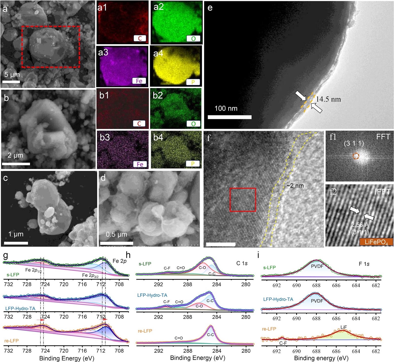
The rapid accumulation of spent LiFePO4 (LFP) cathodes from retired lithium-ion batteries necessitates the development of effective and environmental-friendly recycling strategies. In this context, direct regeneration has emerged as a promising approach for reclaiming LFP cathode materials, offering a streamlined pathway to restore their electrochemical functionality. We report an integrated regeneration protocol that simultaneously repairs the degraded crystal structure and reconstructs the damaged carbon coating in spent LFP. The regenerated cathode material had superfast lithium-ion diffusion kinetics and a stable cathode–electrolyte interface, giving a remarkable rate capability with specific capacities of 122 mAh g−1 at 5C and 106 mAh g−1 at 10C (1C = 170 mA g−1). It also maintained capacities of 110.7 mAh g−1 (5C) and 84.1 mAh g−1 (10C) after 400 cycles. It could be used in harsh environments and could be stably cycled at subzero temperatures (−10 and −20 °C) and in solid-state electrolyte batteries. Life cycle assessment combined with economic evaluation using the EverBatt model reveals that this direct regeneration approach has high economic and environmental benefits.
Oxide Semiconductor for Advanced Memory Architectures: Atomic Layer Deposition, Key Requirement and Challenges
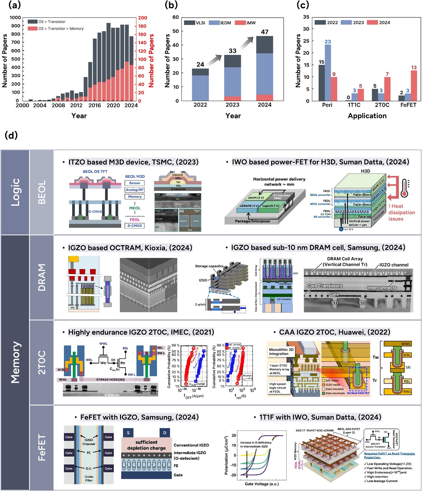
Oxide semiconductors (OSs), introduced by the Hosono group in the early 2000s, have evolved from display backplane materials to promising candidates for advanced memory and logic devices. The exceptionally low leakage current of OSs and compatibility with three-dimensional (3D) architectures have recently sparked renewed interest in their use in semiconductor applications. This review begins by exploring the unique material properties of OSs, which fundamentally originate from their distinct electronic band structure. Subsequently, we focus on atomic layer deposition (ALD), a core technique for growing excellent OS films, covering both basic and advanced processes compatible with 3D scaling. The basic surface reaction mechanisms—adsorption and reaction—and their roles in film growth are introduced. Furthermore, material design strategies, such as cation selection, crystallinity control, anion doping, and heterostructure engineering, are discussed. We also highlight challenges in memory applications, including contact resistance, hydrogen instability, and lack of p-type materials, and discuss the feasibility of ALD-grown OSs as potential solutions. Lastly, we provide an outlook on the role of ALD-grown OSs in memory technologies. This review bridges material fundamentals and device-level requirements, offering a comprehensive perspective on the potential of ALD-driven OSs for next-generation semiconductor memory devices.
Laser powder bed fusion of biodegradable Zn-4Cu alloy: Processing, microstructure and properties
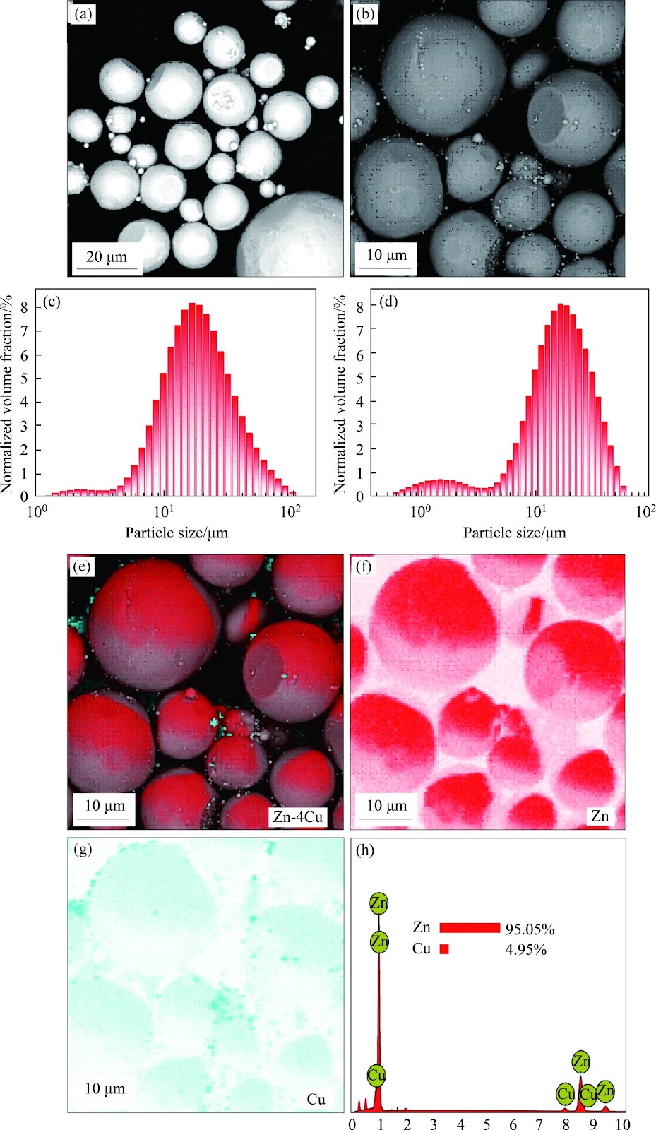
Zn's natural degradability and biocompatibility make it a promising candidate for implants, however, its mechanical properties remain insufficient for bone applications. In this study, the performance of Zn was enhanced by developing Zn-Cu alloys via laser powder bed fusion (LPBF). Optimal LPBF parameters for forming stable tracks were achieved by adjusting laser power and scanning speed. Under optimized conditions of 100 W and 100 mm/s, high-density (99.58%) Zn-Cu alloys with improved hardness (68.2HV) and yield strength (160 MPa) were achieved. These improvements are attributed to solid solution strengthening, segregation strengthening, and grain refinement. The Zn-Cu alloys also demonstrated favorable degradation behavior, with a rate of 0.16 mm/year. This degradation is primarily driven by micro-galvanic corrosion between the CuZn5 phase and Zn matrix, along with refined grains and increased grain boundary density. This work demonstrates a viable strategy for fabricating Zn-based implants with enhanced structural integrity and mechanical performance via LPBF.